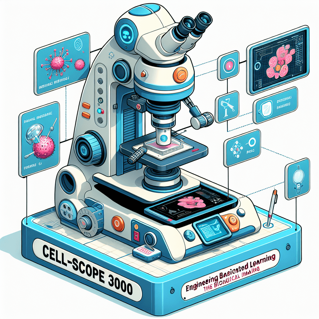
Cell-Scope 3000: Engineering the Future of Biological Imaging
Inquiry Framework
Question Framework
Driving Question
The overarching question that guides the entire project.How can we, as future bio-engineers, design a next-generation imaging tool that overcomes current physical limits to reveal the 4D world of cells and revolutionize how we understand and treat disease?Essential Questions
Supporting questions that break down major concepts.- How have historical breakthroughs in microscopy fueled the evolution of the Cell Theory?
- What are the current technological limitations in imaging, and why do scientists need to see organelles in 4D?
- How does the development of new scientific tools change the way we define and understand "life"?
- In what ways can we apply engineering design principles to overcome the physical barriers of light and scale?
- How would real-time, high-resolution visualization of cell processes impact the future of medicine and disease treatment?
Standards & Learning Goals
Learning Goals
By the end of this project, students will be able to:- Students will analyze the historical progression of microscopy and evaluate how specific technological innovations directly contributed to the development and refinement of Cell Theory.
- Students will identify current physical and technological limitations in cellular imaging (e.g., resolution limits, light toxicity) and explain the scientific necessity of 4D observation for understanding organelle function.
- Students will apply engineering design principles to define a specific imaging problem and conceptualize a tool (the 'Cell-Scope 3000') that utilizes theoretical or emerging technologies to overcome those barriers.
- Students will construct an evidence-based argument describing how real-time, high-resolution visualization of cellular processes could revolutionize medical treatments and our understanding of human health.
Next Generation Science Standards (NGSS)
Common Core State Standards (ELA/Literacy)
Teacher-Provided Standards
Entry Events
Events that will be used to introduce the project to studentsThe Invisible Masterpiece Challenge
Students enter a 'gallery of the unseen' where high-resolution but static images of cells are displayed next to 'lost data' plaques. The teacher reveals that while we can see the 'house' (the cell), we are still blind to the 'party' happening inside in real-time, challenging students to bridge the gap between 3D snapshots and 4D reality.Portfolio Activities
Portfolio Activities
These activities progressively build towards your learning goals, with each submission contributing to the student's final portfolio.The Microscopy Time-Machine
Before students can engineer the future, they must understand the past. In this activity, students act as 'Science Historians' to investigate the timeline of microscopy. They will explore how every leap in cell theory (e.g., discovering the nucleus, identifying organelles) was preceded by a leap in imaging technology—from Leeuwenhoek’s hand-held lenses to modern Electron Microscopes.Steps
Here is some basic scaffolding to help students complete the activity.Final Product
What students will submit as the final product of the activityAn 'Evolution of Vision' Interactive Timeline (digital or physical) that maps at least five major technological innovations to the specific biological discoveries they enabled.Alignment
How this activity aligns with the learning objectives & standardsThis activity aligns directly with Teacher-Specified Standard 1 (Identifying major events and technological innovations) and MS-LS1-1. By tracing the history of the microscope, students understand that scientific knowledge of cells is inextricably linked to the technology available to observe them.The 4D Organelle Mission Brief
Students transition from historians to 'Biological Detectives.' They will choose one specific cellular process (like mitosis, protein transport, or ATP production) and analyze what current static 3D imaging 'misses.' They will define why seeing this process in 4D (real-time movement) is essential for modern medicine and understanding life.Steps
Here is some basic scaffolding to help students complete the activity.Final Product
What students will submit as the final product of the activityAn 'Organelle Action Dossier' that includes a detailed diagram of a chosen organelle and a written 'Gap Analysis' explaining what we still cannot see about its real-time behavior.Alignment
How this activity aligns with the learning objectives & standardsThis activity aligns with MS-LS1-2 (Functions of cell parts) and MS-ETS1-1 (Defining the problem). By focusing on the 'missing 4th dimension' (time), students identify the specific biological gaps that their future engineering project needs to fill.Breaking the Light Barrier: Design Specs
Now acting as 'Bio-Engineers,' students will define the technical requirements for the Cell-Scope 3000. They will research the 'Enemies of Imaging': the Diffraction Limit (light blurring), Phototoxicity (lasers killing the cell), and Resolution. They will create a set of 'Design Constraints' that their conceptual tool must overcome.Steps
Here is some basic scaffolding to help students complete the activity.Final Product
What students will submit as the final product of the activityA 'Cell-Scope 3000 Technical Spec Sheet' listing the target resolution, the method for keeping cells alive (minimizing toxicity), and the proposed 'Future Tech' (e.g., AI-enhancement, quantum sensors) they plan to use.Alignment
How this activity aligns with the learning objectives & standardsThis activity aligns with MS-ETS1-1 (Criteria and constraints) and RST.6-8.7 (Integrating technical information). Students must grapple with the physical 'rules' of science, such as the diffraction limit of light and phototoxicity, to define the boundaries of their engineering solution.Blueprint for the Unseen: The Cell-Scope 3000
In the final phase, students create the 'Cell-Scope 3000.' This is a conceptual blueprint and pitch for their imaginary imaging tool. They must visualize the tool and, more importantly, visualize the 4D output: what does a cell look like through this lens? They will conclude by arguing how this tool would change the future of medicine (e.g., watching a virus enter a cell in real-time).Steps
Here is some basic scaffolding to help students complete the activity.Final Product
What students will submit as the final product of the activityA 'Cell-Scope 3000 Portfolio' consisting of a detailed labeled blueprint of the tool and a '4D Simulation Storyboard' showing four frames of a cellular process as seen through their invention.Alignment
How this activity aligns with the learning objectives & standardsThis activity aligns with MS-ETS1-1 (Developing a solution), MS-LS1-1 (Evidence of cell life), and RST.6-8.7 (Visual models). It serves as the project's capstone, combining biological knowledge with engineering design and communication.Rubric & Reflection
Portfolio Rubric
Grading criteria for assessing the overall project portfolioCell-Scope 3000: Engineering the Future of Cell Biology
Scientific Foundations & History
Evaluates the student's understanding of how microscopy history and technological leaps have directly fueled the evolution of the Cell Theory.Historical & Technical Evolution
The ability to identify and analyze the relationship between historical technological innovations and the resulting advancements in cell theory.
Exemplary
4 PointsPrecisely identifies five or more pivotal innovations, providing a sophisticated analysis of how each specific tool fundamentally shifted scientific paradigms and enabled specific biological breakthroughs. Synthesis of cause-and-effect is exceptionally clear.
Proficient
3 PointsIdentifies five pivotal innovations and accurately describes the scientific breakthrough each allowed. Demonstrates a clear cause-and-effect relationship between the tool and the resulting knowledge of cell biology.
Developing
2 PointsIdentifies three to four innovations but the connection to specific biological breakthroughs is inconsistent or lacks detail. The cause-and-effect relationship is mentioned but not fully developed.
Beginning
1 PointsIdentifies fewer than three innovations or provides a simple list without explaining how these tools led to new scientific understanding. Connections are missing or inaccurate.
Biological Analysis & Application
Assesses the student's capacity to transition from scientific history to current biological needs, specifically focusing on the 4th dimension (time).Organelle Analysis & Gap Identification
The ability to analyze an organelle's function and identify the specific limitations of current 3D static imaging compared to the needs of 4D real-time observation.
Exemplary
4 PointsProvides a profound analysis of a chosen organelle, identifying subtle 'blind spots' in current imaging with high precision. Mission statement reflects a deep understanding of temporal biological processes.
Proficient
3 PointsCorrectly identifies an organelle and its function. Clearly explains the 'Gap Analysis' between static 3D images and 4D real-time needs, resulting in a focused Mission Statement.
Developing
2 PointsIdentifies an organelle but the 'Gap Analysis' is vague. The mission statement is broad and does not clearly specify what behavior or scale the tool needs to capture.
Beginning
1 PointsFails to identify a specific organelle or provides an inaccurate description of its function. Does not identify the difference between static and 4D imaging.
Engineering Design & Specs
Evaluates the student's ability to apply engineering design principles and grapple with the physical 'rules' of science.Technical Constraints & Problem Solving
Demonstrates an understanding of physical limits (diffraction, phototoxicity) and the ability to define engineering constraints and innovative solutions.
Exemplary
4 PointsMasterfully integrates technical concepts like the diffraction limit and phototoxicity into a comprehensive set of constraints. Proposed 'Future Tech' solutions are innovative, grounded in emerging science, and highly creative.
Proficient
3 PointsAccurately explains the diffraction limit and phototoxicity. Lists logical engineering constraints and proposes viable emerging technologies to bypass these physical barriers.
Developing
2 PointsShows a basic understanding of light limits and phototoxicity but struggles to translate them into specific design constraints. Proposed solutions lack technical detail or feasibility.
Beginning
1 PointsDemonstrates significant misconceptions about the physical limits of light or phototoxicity. Design constraints are missing or irrelevant to the identified problems.
Synthesis & Communication
Focuses on the final synthesis of the project: creating the conceptual tool and communicating its value to society.Conceptual Modeling & Visualization
The quality of the conceptual blueprint and the ability to visualize cellular processes across time through a 4D storyboard.
Exemplary
4 PointsProduces a professional-grade labeled blueprint and an exceptional 4-frame storyboard that vividly and accurately depicts complex real-time cellular interactions. Integration of technical specs is seamless.
Proficient
3 PointsCreates a detailed, labeled blueprint of the Cell-Scope 3000 and a 4-frame storyboard that clearly illustrates a logical sequence of an organelle process in real-time.
Developing
2 PointsThe blueprint is missing key labels or technical components. The storyboard shows basic movement but lacks the '4D' detail or sequential clarity required for the project.
Beginning
1 PointsThe model or blueprint is incomplete or messy. The storyboard fails to show a sequential process or does not reflect the capabilities of the designed tool.
Impact Pitch & Reflection
The ability to articulate the potential impact of new technology on medicine and reflect on the changing nature of scientific understanding.
Exemplary
4 PointsDelivers a compelling, evidence-based pitch linking the tool to specific medical breakthroughs. Reflection shows deep metacognition regarding how tools define our understanding of life.
Proficient
3 PointsProvides a clear 'Impact Pitch' explaining how the tool could treat a specific disease. Reflection accurately connects the tool to the historical timeline and the definition of understanding.
Developing
2 PointsThe impact pitch is generic (e.g., 'it helps cure cancer') without explaining the 'how'. Reflection is brief and does not fully address the historical or philosophical connection.
Beginning
1 PointsMinimal or missing impact pitch and reflection. Fails to explain why seeing in 4D matters for the future of medicine or science.