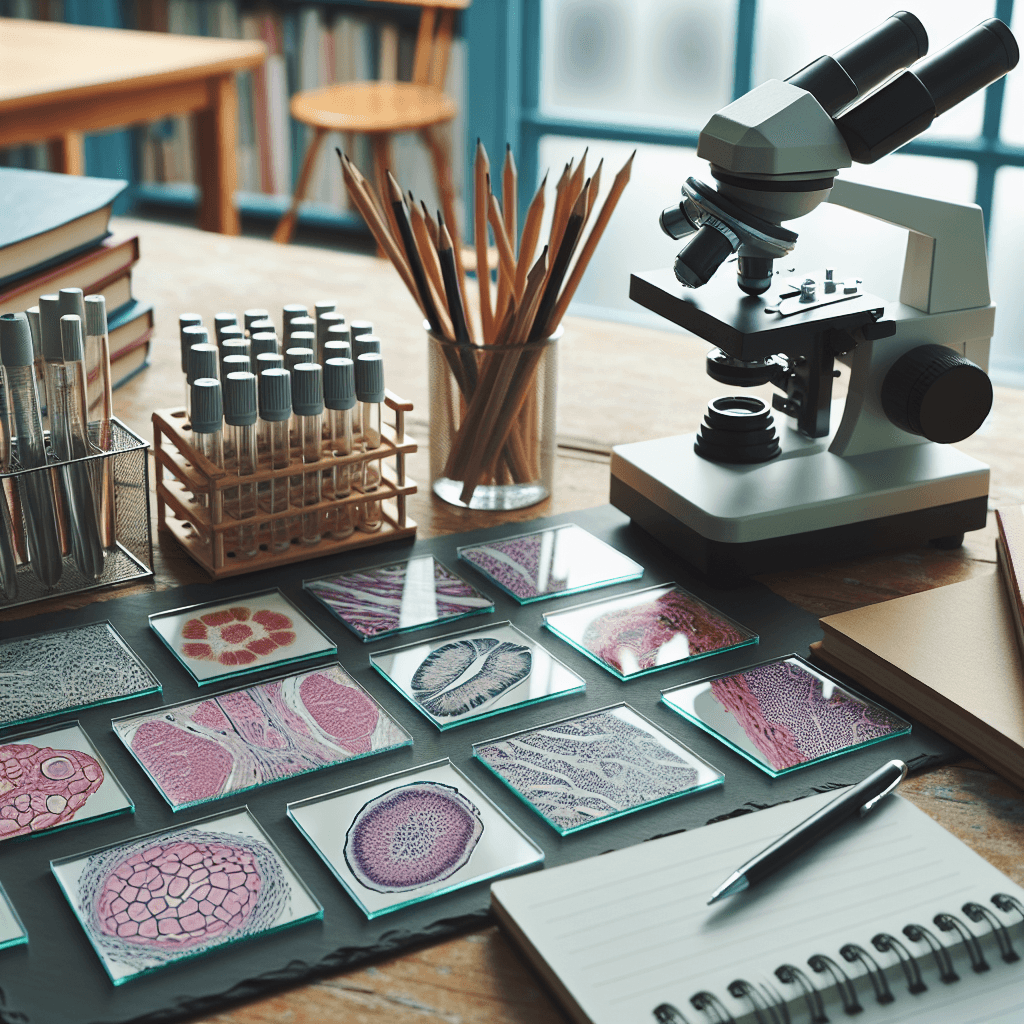
Histology Slide Identification Project
Inquiry Framework
Question Framework
Driving Question
The overarching question that guides the entire project.How can we, as undergraduate students, create a comprehensive guide that uses microscopy to accurately identify and differentiate the diverse types of human tissues, while addressing ethical considerations in tissue usage?Essential Questions
Supporting questions that break down major concepts.- What are the four basic types of tissues in the human body, and how do their structures relate to their functions?
- How can we differentiate between different types of epithelial tissue based on their cellular arrangements and specializations?
- What are the main components of connective tissue, and how do they contribute to its diverse roles in the body?
- How do muscle tissues differ in terms of their cellular structure, mechanism of contraction, and control?
- What are the key features of nervous tissue, and how do they enable communication within the body?
- How can we use microscopy techniques to visualize and identify different tissue types and cellular structures?
- What are the common artifacts encountered in histological slides, and how can we distinguish them from genuine tissue features?
- How does the organization of cells and extracellular matrix contribute to the overall function of organs and systems?
- What are the ethical considerations in using human tissues for educational and research purposes?
Standards & Learning Goals
Learning Goals
By the end of this project, students will be able to:- Students will be able to identify and differentiate the four basic tissue types (epithelial, connective, muscle, and nervous) under a microscope.
- Students will be able to relate the structure of different tissues to their functions within the human body.
- Students will be able to prepare and stain histological slides to visualize tissue structures.
- Students will be able to distinguish between normal and pathological tissue samples.
- Students will be able to understand and address the ethical considerations related to the use of human tissues in histology.
Entry Events
Events that will be used to introduce the project to studentsHistological "CSI" Scenario
**Histological 'CSI' Scenario:** Students are presented with a mock 'crime scene' where the key evidence is a tissue sample. They must use their histology knowledge to analyze the slide, determine the cause of 'death' or 'failure', and present their findings in a formal 'expert witness' report, connecting histology to real-world problem-solving.Portfolio Activities
Portfolio Activities
These activities progressively build towards your learning goals, with each submission contributing to the student's final portfolio.Structure-Function Spotlight
Students research and present on the structure-function relationship of a specific tissue type. This activity emphasizes the connection between microscopic anatomy and physiological roles.Steps
Here is some basic scaffolding to help students complete the activity.Final Product
What students will submit as the final product of the activityA short presentation (PowerPoint, poster, or oral) explaining how the structure of a chosen tissue type enables it to perform its specific functions.Alignment
How this activity aligns with the learning objectives & standardsLearning Goal: Students will be able to relate the structure of different tissues to their functions within the human body.Rubric & Reflection
Portfolio Rubric
Grading criteria for assessing the overall project portfolioHistology Structure-Function Rubric
Tissue Structure-Function Presentation
Focuses on evaluating the student's ability to accurately identify tissue structures, explain structure-function relationships, and present findings effectively.Structural Identification Accuracy
Accuracy of the identified structures and components within the chosen tissue type.
Exemplary
4 PointsDemonstrates comprehensive and accurate identification of all key structures and components of the tissue, including cellular and extracellular elements. Information presented is detailed and precise.
Proficient
3 PointsDemonstrates accurate identification of most key structures and components of the tissue. Minor inaccuracies or omissions may be present but do not detract from overall understanding.
Developing
2 PointsDemonstrates emerging identification skills with some correct structures and components identified, but significant inaccuracies or omissions are evident.
Beginning
1 PointsDemonstrates limited ability to identify structures and components of the tissue. Significant inaccuracies and omissions are prevalent.
Structure-Function Explanation
Clarity and depth of the explanation of the relationship between the tissue's structure and its functions.
Exemplary
4 PointsProvides an exceptionally clear, detailed, and insightful explanation of how the tissue's structure enables its specific functions, demonstrating a deep understanding of structure-function relationships at a microscopic level. Connects structure to function in a novel or sophisticated way.
Proficient
3 PointsProvides a clear and thorough explanation of how the tissue's structure enables its specific functions. Demonstrates a strong understanding of structure-function relationships.
Developing
2 PointsProvides an explanation of the relationship between structure and function, but the explanation is superficial, incomplete, or contains some inaccuracies.
Beginning
1 PointsProvides a limited or unclear explanation of how the tissue's structure relates to its function. Demonstrates a weak understanding of structure-function relationships.
Visual Aid Quality
Effectiveness and appropriateness of visual aids (micrographs, diagrams) in supporting the presentation.
Exemplary
4 PointsVisual aids are exceptionally clear, relevant, and effectively enhance understanding of the tissue's structure and function. Visuals are high-quality, accurately labeled, and creatively presented to maximize learning impact.
Proficient
3 PointsVisual aids are clear, relevant, and effectively support understanding of the tissue's structure and function. Visuals are accurately labeled and contribute to overall clarity.
Developing
2 PointsVisual aids are present but may be unclear, not entirely relevant, or only partially support understanding of the tissue's structure and function. Labeling may be incomplete or inaccurate.
Beginning
1 PointsVisual aids are missing, irrelevant, or do not support understanding of the tissue's structure and function. Visuals may be poorly presented or inaccurate.
Presentation Quality
Overall presentation quality, including organization, clarity, and engagement.
Exemplary
4 PointsPresentation is exceptionally well-organized, clear, and engaging. The presenter demonstrates a deep understanding of the material and communicates effectively with the audience, fostering interest and understanding. Demonstrates leadership in presentation quality.
Proficient
3 PointsPresentation is well-organized, clear, and engaging. The presenter demonstrates a good understanding of the material and communicates effectively with the audience.
Developing
2 PointsPresentation is somewhat organized but may lack clarity or engagement. The presenter demonstrates a basic understanding of the material, but communication may be inconsistent.
Beginning
1 PointsPresentation is poorly organized, unclear, and unengaging. The presenter demonstrates a limited understanding of the material and struggles to communicate effectively.